The rising incidence of non-endemic, tissue-destructive parasites in domestic pets represents a structural failure in international veterinary biosecurity, shifting biological risk from isolated rural livestock operations directly into suburban households. When popular media warns of "flesh-eating parasites" lurking on companion animals, they are typically conflating two distinct biological threats: Leishmania infantum, a protozoan transmitted via phlebotomine sandflies that causes severe visceral and cutaneous lesions, and Cochliomyia hominivorax (the New World screwworm), an obligate parasite whose larvae consume living mammalian tissue. Managing this veterinary risk requires abandoning emotional panic in favor of structured epidemiological screening, mechanical threat identification, and strict vector-management protocols.
Understanding the threat vectors requires analyzing the specific biological mechanisms by which these organisms colonize a host. Pet owners and veterinary practitioners frequently misdiagnose early-stage tissue parasitism because the initial clinical presentations mimic benign dermatological conditions like allergic dermatitis, hot spots, or standard pyoderma. Failing to differentiate these conditions creates an diagnostic bottleneck, allowing the infestation or infection to progress to irreversible systemic failure or severe localized necrosis.
The Two Vectors of Tissue-Destructive Parasitism
To systematically defend a domestic environment against these organisms, one must categorize them by their primary pathogenic mechanism: intracellular systemic colonization or extracellular necrotic infestation.
Intracellular Systemic Colonization: Leishmania infantum
Leishmania infantum does not chew tissue from the outside; instead, it hijacks the host’s immune response. The parasite enters the mammalian host through the bite of an infected female sandfly. Once inside the skin, the flagellated promastigote form is engulfed by macrophages—the very immune cells sent to destroy it. Instead of being digested, the parasite transforms into an unflagellated amastigote, multiplying within the phagolysosome until the host cell ruptures.
This cellular destruction creates a cascade of systemic failure. The clinical manifestations follow a predictable sequence:
- Exfoliative Dermatitis: A dry, flaky coat accompanied by hair loss, typically starting around the eyes (periorbital alopecia) and muzzle. This is often mistaken for seasonal allergies.
- Hyperkeratosis: Thickening and cracking of the skin at pressure points, such as the elbows and footpads.
- Onychogryphosis: Abnormal, accelerated nail growth caused by chronic inflammation of the nail bed, resulting in long, curved, fragile claws.
- Visceral Dissemination: The parasite migrates to internal organs, primarily the spleen, liver, and kidneys. Immune complex deposition in the renal glomeruli causes immune-mediated glomerulonephritis, leading to chronic renal failure, which is the primary cause of mortality in infected canines.
Extracellular Necrotic Infestation: Cochliomyia hominivorax
Unlike Leishmania, the New World screwworm operates through direct, mechanical tissue consumption. The female fly is attracted to any open wound, no matter how minor—including tick bites, surgical incisions, or the umbilicus of newborn animals. She deposits up to 400 eggs along the margins of the wound.
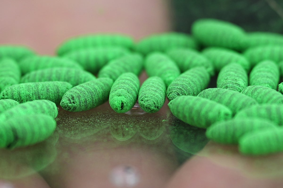
Within 12 to 24 hours, the larvae hatch and immediately bore into the living tissue. The structural breakdown of this infestation follows an aggressive timeline:
- Lytic Destruction: The larvae secrete proteolytic enzymes that liquefy the surrounding muscle and dermal tissue, allowing them to feed exclusively on living flesh. This differentiates them from common blowfly maggots, which primary consume necrotic or dead tissue.
- Mechanical Cavitation: As the larvae feed, they burrow deeper into the host, creating a pocket or cavity beneath the skin that remains largely hidden from view, save for a small opening used for respiration.
- Secondary Toxemia: The excretion of metabolic waste products by hundreds of larvae inside the living tissue cavity induces systemic toxemia and septicemia. Without intervention, the host experience septic shock within several days of hatching.
Diagnostic Frameworks for Veterinary Screening
Differentiating these parasitic threats from standard domestic infections requires an objective diagnostic framework. Relying on visual inspection alone introduces an unacceptable margin of error.
                  [Suspected Lesion / Dermal Anomaly]
                                   |
         -----------------------------------------------------
        |                                                     |
[Exudative Wound w/ Movement]                 [Dry, Flaky Lesion / Alopecia]
        |                                                     |
 (Screwworm Protocol)                              (Leishmania Protocol)
        |                                                     |
1. Microscopic Larval Assessment             1. Quantitative Serology (ELISA)
2. Verify Posterior Spiracles                2. PCR Testing (Tissue Aspirate)
Protocol for Suspected Screwworm Infestation
When a pet presents with an exudative, foul-smelling wound, the immediate objective is to identify whether the infesting larvae are obligate parasites (Cochliomyia hominivorax) or facultative scavengers (common blowflies).
The practitioner must extract several larvae from the deep margins of the wound using forceps. Place the larvae in a preservative solution and examine them under a stereomicroscope. The defining anatomical feature of the New World screwworm is the presence of dark, heavily pigmented tracheal trunks extending anteriorly from the posterior spiracles (breathing pores) along the dorsal surface of the last three body segments. If these dark lines are visible, the case must be reported to state or national agricultural authorities immediately, as screwworm is a highly regulated, reportable agricultural pest.
Protocol for Suspected Leishmaniasis
Because Leishmania is an intracellular pathogen, identification requires evaluating cellular and serological markers.
Quantitative serology via Enzyme-Linked Immunosorbent Assay (ELISA) or Indirect Immunofluorescent Antibody Test (IFAT) serves as the first line of screening. High antibody titers correlate strongly with active, symptomatic disease. However, subclinically infected dogs can yield borderline or negative results during the long incubation period, which ranges from months to years.
For a definitive diagnosis, Polymerase Chain Reaction (PCR) assays offer the highest sensitivity and specificity. Sampling should target tissues with a high parasite load. Bone marrow or lymph node aspirates yield the highest diagnostic accuracy, followed by skin scrapings or biopsies taken from the margins of active ulcers. Quantitative PCR (qPCR) is preferred, as it measures the parasite load, providing a baseline to evaluate treatment efficacy.
Prophylaxis and Environmental Risk Mitigation
Eradicating these parasites once they have established a foothold in a canine host is difficult, expensive, and in the case of Leishmania, rarely results in a complete parasitological cure. Prevention relies on breaking the vector-host transmission cycle through chemical and physical barriers.
Chemical Vector Repellents
Standard flea and tick preventatives (such as isoxazolines) are insufficient against phlebotomine sandflies. Protection against Leishmania requires topical formulations containing permethrin or deltamethrin. These synthetic pyrethroids provide an anti-feeding effect, preventing the sandfly from biting and transferring the promastigotes. These compounds must be administered on a strict, continuous schedule throughout the active vector season, which expands as global temperatures rise.
Environmental Engineering
Sandflies are weak fliers and are most active between dusk and dawn. They thrive in microclimates with high humidity and organic debris, such as leaf litter, compost piles, and animal shelters.
- Microclimate Destruction: Remove decaying organic matter within a 50-meter radius of the domestic perimeter to eliminate sandfly breeding sites.
- Structural Barriers: Keep pets indoors during peak feeding hours (dusk to dawn). Standard window screens do not block sandflies due to their minute size (1.5 to 3 mm); fine-mesh netting treated with insecticide is required for kennels or open windows.
- Wound Management: For screwworm prevention, any open wound on an animal must be treated immediately with a topical larvicide (such as formulations containing spinosad or ivermectin) and kept clean until fully healed. Pets should be monitored closely following outdoor excursions in endemic or transitional geographic zones.
Treatment Protocols and Biological Limitations
When prevention fails, the therapeutic options are constrained by biological realities and pharmaceutical availability.
Managing Leishmaniasis
There is no universally available vaccine that provides sterilizing immunity against Leishmania infantum. Treatment focuses on reducing parasite burden and managing clinical symptoms. The standard first-line regimen in endemic regions combines meglumine antimoniate (a leishmanicidal agent) with allopurinol (a leishmanistatic agent). Meglumine antimoniate disrupts the parasite's energy metabolism, while allopurinol inhibits protein synthesis by acting as a purine analogue.
The primary limitation of this strategy is that meglumine antimoniate carries a high risk of nephrotoxicity, which can exacerbate pre-existing renal damage caused by the disease itself. Furthermore, while clinical remission is common, the parasite persists in low numbers within the bone marrow and lymphoid organs, leading to eventual relapses that require life-long monitoring and repeated courses of therapy.
Managing Screwworm Infestation
The therapeutic approach to Cochliomyia hominivorax is mechanical and surgical rather than purely pharmaceutical.
The primary step involves the physical removal of all visible larvae from the wound cavity using surgical instruments. This must be followed by thorough debridement of the wound to remove all necrotic, liquefied tissue that could otherwise promote secondary bacterial infections.
Following debridement, apply a topical insecticide directly into the cavity to destroy any remaining microscopic or deeply embedded larvae. Administer systemic antibiotics (such as amoxicillin-clavulanate or cephalexin) to treat the secondary bacterial pyoderma that inevitably accompanies a screwworm infestation. The main limitation here is the risk of missing larvae that have migrated deep into muscular tissue or body cavities, which can cause internal necrosis and systemic sepsis even after the primary surface wound appears to be clearing.
Geographic Range Shifts and Biosecurity Vulnerabilities
The proliferation of these tissue parasites is accelerated by international pet rescue travel and shifting climate zones. Sandflies are migrating northward into previously temperate regions due to rising average winter temperatures, allowing Leishmania to establish endemic cycles in areas previously considered safe. Concurrently, the importation of rescue dogs from endemic regions without rigorous screening introduces a reservoir of infected hosts, bypassing traditional geographical barriers.
Vigilance requires a shift from reactive treatment to proactive, multi-tiered screening. Pet owners traveling through or importing animals from endemic zones must implement pre-travel PCR testing, continuous pyrethroid application, and immediate isolation of any animal presenting with non-responsive cutaneous lesions. Relying on basic hygiene or standard flea collars is an obsolete strategy against specialized, tissue-destructive parasites.